NEWS
New bioprinting method improves the development of realistic tumour models
Researchers at the Research Campus STIMULATE, in collaboration with other partners at Otto von Guericke University Magdeburg, have developed a new method that enables tumour models to be produced with exceptional precision in the laboratory. The scientific paper, entitled “Precision bioprinting-based extrusion of tumour spheroids on pre-matured in vitro tissue models on demand”, has been accepted for publication in the high-impact journal IOP Biofabrication (Impact Factor: 9.3). The first authors of the publication are Marieke Kramer (Core Facility Tissue Engineering) and Klaudija Janic (Organic Chemistry Group led by Prof. Julian Thiele) (shared first authorship); the corresponding author is Dr Sascha Kopp, head of the Biophantoms research group at the Research Campus STIMULATE and the Core Facility Tissue Engineering.
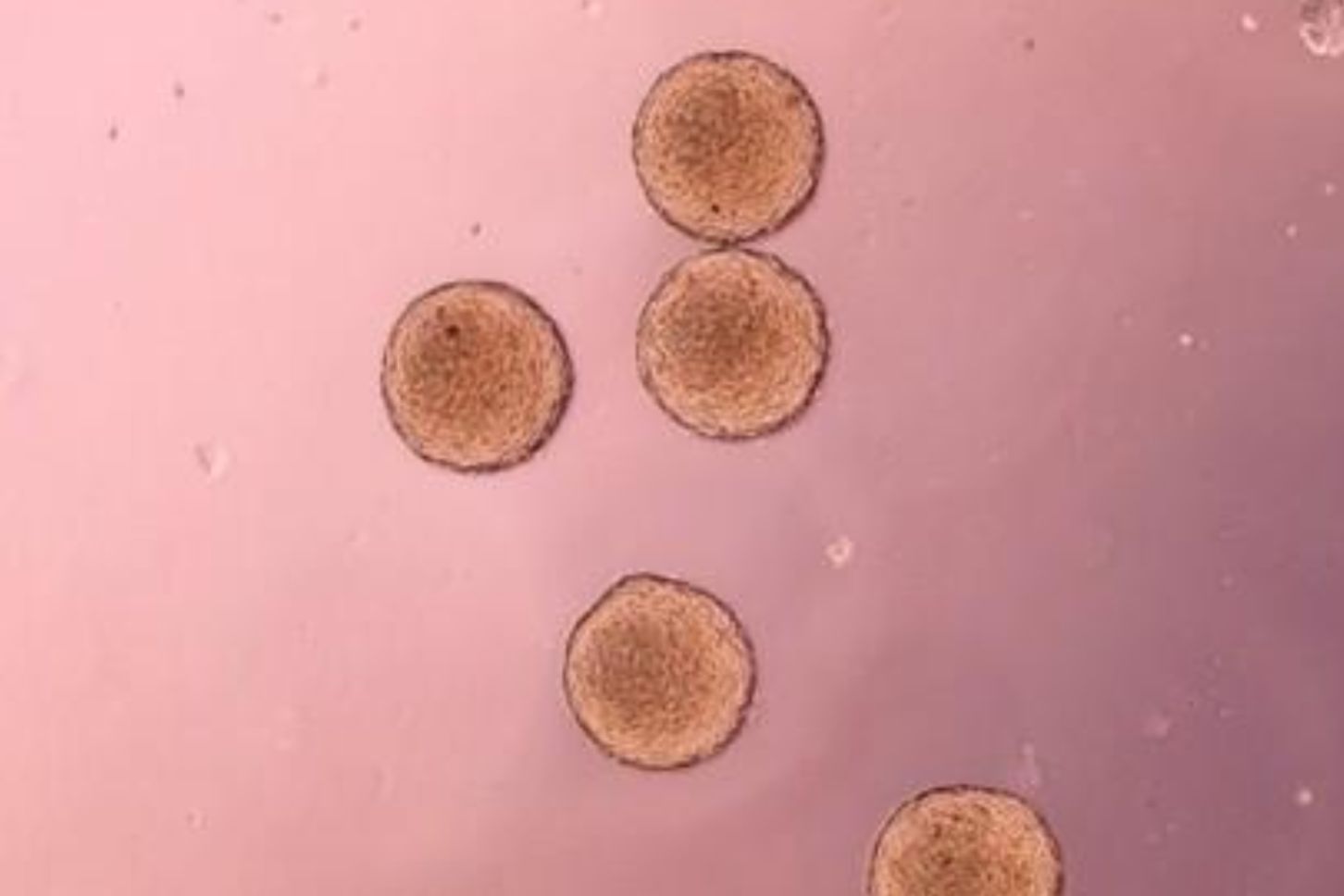
The focus of the research was on a specially adapted 3D bioprinting process that enables tiny clusters of tumour cells (tumour spheroids) to be placed precisely on artificially produced tissue models. To this end, the researchers developed, amongst other things, a new holder for the tissue models, custom-programmed software for precise positioning, and a standardised printing process. This enables tumour models to be produced reproducibly and with a high degree of precision.
Such realistic tissue models make it possible to test and further develop new imaging methods and minimally invasive diagnostic and therapeutic technologies under standardised conditions. In the long term, they can help to bring medical innovations into clinical use more quickly, whilst reducing the need for animal testing.
With this work, the Research Campus STIMULATE demonstrates its expertise at the interface between bioprinting, tissue models and image-guided medical technology. The methods developed lay an important foundation for the development and validation of innovative technologies for cancer diagnosis and treatment. The research was funded by the European Regional Development Fund (ERDF) as part of the Saxony-Anhalt Science Priorities initiative.
The original publication can be viewed here.
Neue Bioprinting-Methode verbessert Entwicklung realistischer Tumormodelle
Forschende des Forschungscampus STIMULATE haben gemeinsam mit weiteren Partnern der Otto-von-Guericke-Universität Magdeburg eine neue Methode entwickelt, mit der sich Tumormodelle im Labor besonders präzise herstellen lassen. Die wissenschaftliche Arbeit mit dem Titel „Precision bioprinting-based extrusion of tumour spheroids on pre-matured in vitro tissue models on demand“ wurde im hochrangigen Fachjournal IOP Biofabrication (Impact Factor: 9,3) angenommen. Erstautorinnen der Publikation sind Marieke Kramer (Core Facility Tissue Engineering) und Klaudija Janic (Arbeitskreis Organische Chemie Prof. Julian Thiele) (geteilte Erstautorenschaft), Letztautor ist Dr. Sascha Kopp, Leiter der Forschungsgruppe Biophantoms am Forschungscampus STIMULATE und der Core Facility Tissue Engineering.
Im Mittelpunkt der Forschung stand ein speziell angepasster 3D-Bioprinting-Prozess, mit dem winzige Tumorzellverbände (Tumorsphäroide) gezielt auf künstlich hergestellten Gewebemodellen platziert werden können. Dafür entwickelten die Forschenden unter anderem eine neue Halterung für die Gewebemodelle, eine eigens programmierte Software zur exakten Positionierung sowie einen standardisierten Druckprozess. So lassen sich Tumormodelle reproduzierbar und mit hoher Präzision herstellen.
Solche realitätsnahen Gewebemodelle ermöglichen es, neue bildgebende Verfahren und minimal-invasive Diagnostik- und Therapietechnologien unter standardisierten Bedingungen zu testen und weiterzuentwickeln. Langfristig können sie dazu beitragen, medizinische Innovationen schneller in die klinische Anwendung zu bringen und gleichzeitig den Bedarf an Tierversuchen zu reduzieren.
Mit der Arbeit unterstreicht der Forschungscampus STIMULATE seine Kompetenz an der Schnittstelle von Bioprinting, Gewebemodellen und bildgeführter Medizintechnik. Die entwickelten Methoden schaffen eine wichtige Grundlage für die Entwicklung und Validierung innovativer Technologien zur Krebsdiagnostik und -therapie. Die Forschungsarbeiten wurden durch den Europäischen Fonds für regionale Entwicklung (EFRE) im Rahmen der Wissenschaftsschwerpunkte Sachsen-Anhalt (Saxony-Anhalt Science Priorities initiative) gefördert.
Die Originalpublikation ist hier einsehbar.