NEWS
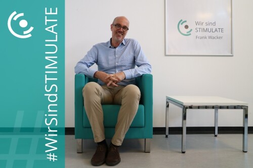
Interview ‘We are STIMULATE’ with Prof Dr Frank Wacker
Interviewee: Prof Dr Frank Wacker
Position: Medical Director of the Oncology/MRI focus area
Interviewer: Lea Nickel
Date: 10.04.2025

You are Director of the Institute of Diagnostic and Interventional Radiology at Hannover Medical School (MHH) and also head the Imaging Unit at the Clinical Research Centre Hannover. What does a typical working week look like for you?
Reply: There isn't really a typical week; every day brings different tasks. On Monday, for example, I was in the clinic, saw patients and then took part in committee and planning meetings. On Tuesday, I wrote a research proposal and went to angiography, and on Wednesday I made phone calls and held video conferences. The constants are the daily early meeting at eight o'clock and the release of findings in the afternoon and evening, everything else varies greatly between research, clinical and management tasks.
Could you give us a brief outline of your professional career?
Reply: I studied medicine in Tübingen and completed my practical year partly in Tübingen and partly in Esslingen. I then went to Berlin for my residency at what was then the Steglitz University Hospital, which later became part of the Charité. I also completed my further training as a radiologist there. My enthusiasm for interventional radiology took hold of me in the third year of my training and continues to this day. From Berlin I went to the USA, to the University Hospital of Case Western Reserve University in Cleveland, where I worked in interventional radiology and did research in the field of interventional MRI, which was also the subject of my habilitation in Berlin. After two years, I returned to Germany, initially working in MR development at Siemens and later again at the Charité, where I took on a C3 professorship. This was followed by further clinical and scientific work in the USA, this time at Johns Hopkins University, before I finally accepted a professorship at the MHH, where I have been working ever since.
Why was it important to you to combine medical practice with scientific research from the outset?
Reply: What fascinated me about studying medicine right from the start was the openness - you can take very different paths, from general practice to university medicine. For me, the enthusiasm for research came early on through my doctoral thesis. Back then, I was analysing meniscus lesions using MRI. This was still a new diagnostic procedure at the time, but its potential immediately grabbed me. My doctoral supervisor then brought me to the University Hospital in Berlin, where it was easy to combine both clinical work and research. It's crucial that you have the inner drive to really want to develop medicine further. This motivation is still with me today.
At STIMULATE, you contribute your expertise as medical director in the fields of oncology and interventional MRI. What do you find particularly appealing about interdisciplinary collaboration in this context?
Reply: My enthusiasm for interdisciplinary collaboration has its origins in my time in Cleveland. There I was part of a small, very committed research group on interventional MRI, with physicists, doctors and doctoral students from a wide range of disciplines. This close collaboration at eye level had a huge impact on me. That's exactly what I wanted to continue in Germany, and I succeeded. At the MHH, we now supervise around 15 doctoral students from the natural sciences. Working at the Research Campus STIMULATE was then a logical extension: an opportunity to contribute our clinical perspective to joint projects with computer scientists, engineers and physicists. These collaborations are incredibly enriching - and work really well here.
In addition to image-guided therapy, your research topics also include machine learning and AI. How is artificial intelligence changing everyday working life in radiology - and what opportunities or limitations do you currently see?
Reply: Radiology is predestined for AI applications because our data has long been completely digital and at the same time the volume of images has increased enormously. Artificial intelligence can help us to analyse the data. However, a complete replacement of medical expertise is not foreseeable in the near future; AI is currently doing even more work. For some diagnostic decisions, a combination of human and machine is often the best solution. Studies show that these dual findings deliver better results. At the same time, we must remain critical: Many AI systems promise more than they deliver and their benefits are not always proven. That's why it's important that we remain involved in research ourselves - not just as users, but as co-creators.
What tips would you give to young people who work or would like to work in radiology and medical technology in the future?
Reply: Choose a path that you really enjoy and be open to change. Many things are developing rapidly in radiology and medical technology, so it's worth thinking ahead: Which roles could become important in 10 or 20 years? Which competences can also be used in other fields? Leadership, organisation and project management are all easily transferable skills. What is important is the willingness to think outside the box.
Thank you very much for the exciting insight and your time!
Click here for an overview of all interviews conducted so far.
Interview „Wir sind STIMULATE“ mit Prof. Dr. Frank Wacker
Interviewter: Prof. Dr. Frank Wacker
Stelle: Medizinischer Leiter des Fokusfelds Onkologie/MRT
Interviewerin: Lea Nickel
Datum: 10.04.2025
Sie sind Direktor des Instituts für Diagnostische und Interventionelle Radiologie an der Medizinischen Hochschule Hannover (MHH) und leiten zusätzlich die Imaging Unit im Clinical Research Center Hannover. Wie sieht eine typische Arbeitswoche bei Ihnen aus?
Antwort: Eine typische Woche gibt es eigentlich nicht, jeder Tag bringt andere Aufgaben mit sich. Am Montag war ich zum Beispiel in der Klinik, habe Patient:innen gesehen und danach an Gremien- und Planungssitzungen teilgenommen. Am Dienstag habe ich an einem Forschungsantrag geschrieben und war in der Angiografie und am Mittwoch habe ich Telefonate und Videokonferenzen geführt. Konstanten sind die tägliche Frühbesprechung um acht Uhr und die Freigabe von Befunden am Nachmittag und Abend, alles andere variiert stark zwischen Forschung, Klinik und Leitungsaufgaben.
Könnten Sie uns kurz Ihren beruflichen Werdegang skizzieren?
Antwort: Ich habe in Tübingen Medizin studiert und mein praktisches Jahr teilweise in Tübingen, teilweise in Esslingen absolviert. Danach bin ich für meine Assistenzzeit nach Berlin ans damalige Universitätsklinikum Steglitz gegangen, das später Teil der Charité wurde. Dort habe ich auch meine Weiterbildung zum Radiologen abgeschlossen. Bereits im dritten Jahr meiner Weiterbildung hat mich die Begeisterung für die interventionelle Radiologie erfasst, das hält bis heute an. Aus Berlin ging ich in die USA, ans University Hospital der Case Western Reserve University in Cleveland, wo ich in der interventionellen Radiologie gearbeitet und im Bereich interventionelle MRT geforscht habe, das war auch Thema meiner Habilitation in Berlin. Nach zwei Jahren kehrte ich nach Deutschland zurück, arbeitete zunächst in der MR-Entwicklung bei Siemens und später wieder an der Charité, wo ich eine C3-Professur übernahm. Es folgte eine weitere klinische und wissenschaftliche Tätigkeit in den USA, diesmal an der Johns-Hopkins-Universität, bevor ich schließlich dem Ruf an die MHH folgte, wo ich seitdem tätig bin.
Warum war es Ihnen wichtig, die ärztliche Tätigkeit von Anfang an auch mit wissenschaftlicher Forschung zu verbinden?
Antwort: Was mich am Medizinstudium von Anfang an fasziniert hat, war die Offenheit – man kann sehr unterschiedliche Wege einschlagen, von der Hausarztpraxis bis zur Hochschulmedizin. Für mich kam die Begeisterung für Forschung früh über meine Doktorarbeit. Damals habe ich Meniskusläsionen im MRT untersucht. Das war damals ein noch junges diagnostisches Verfahren, dessen Potenzial mich sofort gepackt hat. Durch meinen Doktorvater bin ich dann an die Uniklinik in Berlin gekommen, und dort war es einfach, beides – klinische Arbeit und Forschung – zu verbinden. Entscheidend ist, dass man den inneren Antrieb hat, Medizin wirklich weiterentwickeln zu wollen. Diese Motivation begleitet mich bis heute.
Bei STIMULATE bringen Sie Ihre Expertise als medizinische Leitung in den Bereichen Onkologie und interventionelles MRT ein. Was reizt Sie an der interdisziplinären Zusammenarbeit in diesem Kontext besonders?
Antwort: Meine Begeisterung für interdisziplinäre Zusammenarbeit hat ihren Ursprung in meiner Zeit in Cleveland. Dort war ich Teil einer kleinen, sehr engagierten Forschungsgruppe zur interventionellen MRT, mit Physiker:innen, Ärzt:innen und Promovierenden unterschiedlichster Fachrichtungen. Diese enge Zusammenarbeit auf Augenhöhe hat mich sehr geprägt. Genau das wollte ich in Deutschland fortsetzen und das ist mir gelungen. An der MHH betreuen wir inzwischen rund 15 Promovierende aus den Naturwissenschaften. Die Mitarbeit am Forschungscampus STIMULATE war dann eine logische Erweiterung: eine Chance, unsere klinische Perspektive in gemeinsame Projekte mit Informatiker:innen, Ingenieur:innen und Physiker:innen einzubringen. Diese Kooperationen sind unglaublich bereichernd – und funktionieren hier wirklich gut.
In Ihren Forschungsthemen sind neben der bildgestützten Therapie auch Maschinelles Lernen und KI ein Teil. Wie verändert künstliche Intelligenz den Arbeitsalltag in der Radiologie – und welche Chancen oder auch Grenzen sehen Sie aktuell?
Antwort: Radiologie ist prädestiniert für KI-Anwendungen, denn unsere Daten sind längst vollständig digital, und gleichzeitig haben die Bildmengen enorm zugenommen. Künstliche Intelligenz kann uns bei der Auswertung entlasten. Aber: Ein vollständiger Ersatz ärztlicher Expertise ist in naher Zukunft nicht absehbar, aktuell macht die KI noch mehr Arbeit. Bei manchen diagnostischen Entscheidungen ist die Kombination aus Mensch und Maschine oft die beste Lösung. Studien zeigen, dass diese Doppelbefunde bessere Ergebnisse liefern. Gleichzeitig müssen wir kritisch bleiben: Viele KI-Systeme versprechen mehr, als sie halten, und ihr Nutzen ist nicht immer belegt. Darum ist es wichtig, dass wir selbst an der Forschung beteiligt bleiben – nicht nur als Anwender:innen, sondern als Mitgestalter:innen.
Welche Tipps würden Sie jungen Menschen mit auf den Weg geben, die im Bereich Radiologie und Medizintechnik arbeiten oder zukünftig arbeiten möchten?
Antwort: Wählen Sie einen Weg, der Ihnen wirklich Freude macht und seien Sie offen für Veränderung. In der Radiologie und Medizintechnik entwickelt sich vieles rasant, also lohnt es sich, vorausschauend zu denken: Welche Rollen könnten in 10 oder 20 Jahren wichtig werden? Welche Kompetenzen lassen sich auch in anderen Feldern nutzen? Führung, Organisation oder Projektmanagement sind alles leicht übertragbare Fähigkeiten. Wichtig ist die Bereitschaft, über den Tellerrand hinauszuschauen.
Vielen Dank für den spannenden Einblick und Ihre Zeit!
Hier geht's zur Übersicht über alle bisher geführten Interviews.